
- ULTRASOUND SCANS
Top Rated Ultrasound Scan Available
Ultrasonography, often referred to as ultrasound, is a medical imaging technique that uses high-frequency sound waves to produce real-time images of the internal structures and organs of the body.
Ultrasonography is a non-invasive diagnostic imaging method that relies on the transmission and reflection of sound waves to create detailed images of various parts of the body. It is commonly used in medicine for both diagnostic and monitoring purposes.

3D/4D Scans
All Pregnancy Scans
- Early Pregnancy Scan
- NT Scan
- 1st Trimester Scan
- Anomaly Scan
- 2nd Trimester Scan
- 3rd Trimester Scan
- Growth Scan
Doppler Scans
- Abdominal doppler
- Carotid
- Renal
- Hepatic
- Peripheral arterial and venous dopplers

Perianal / Fistula Ultrasound
Perianal or fistula ultrasound is a specialized medical imaging technique that is used to evaluate perianal and perirectal regions for the presence of fistulas, abscesses, or other abnormalities.
Perianal ultrasound is primarily employed to diagnose and assess anal fistulas. Fistulas are abnormal passages or tunnels that form between the anal canal or rectum and the skin near the anus. They can be painful and often result from underlying conditions such as anal abscesses or Crohn’s disease.
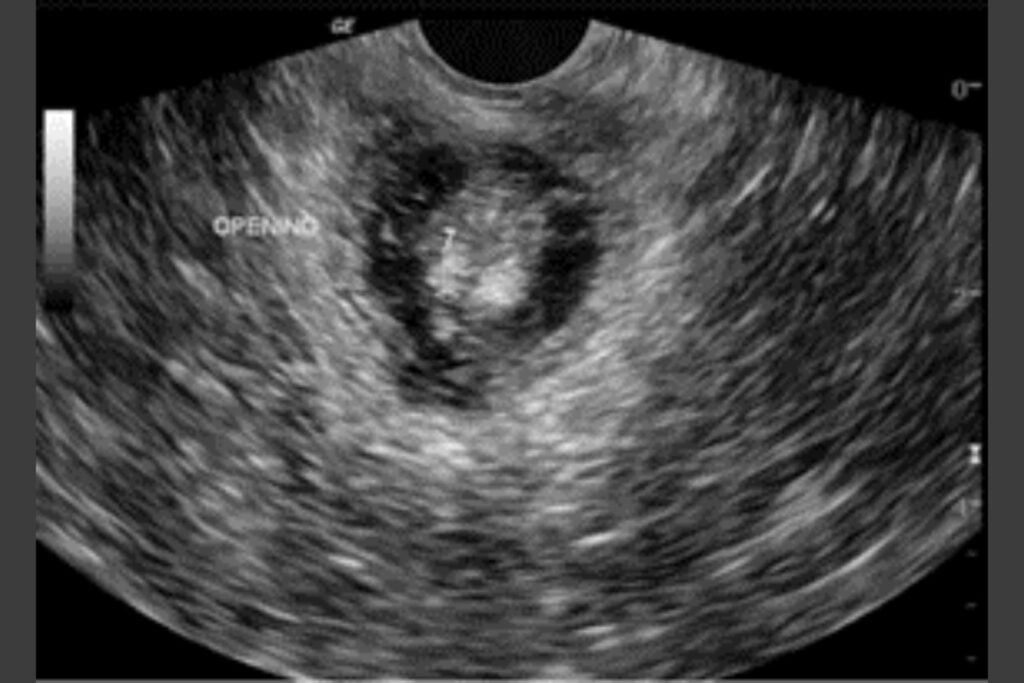
Fetal Neurosonogram


Musculoskeletal Ultrasound and Guided Interventions
Ultrasound Guided Diagnostic Interventions
Ultrasound-guided diagnostic interventions refer to medical procedures in which real-time ultrasound imaging is used to guide the placement of instruments or needles for diagnostic purposes.
These interventions allow healthcare providers to visualize the target area in the body while performing a procedure, increasing accuracy and reducing the risk of complications.


Small Parts Sonography
Small parts sonography, also known as small parts ultrasound, is a specialized medical imaging technique that uses ultrasound technology to visualize and assess various small anatomical structures in the body.
Typically used to examine structures that are not part of the major organs :
- Thyroid
- Breast
- Scrotum
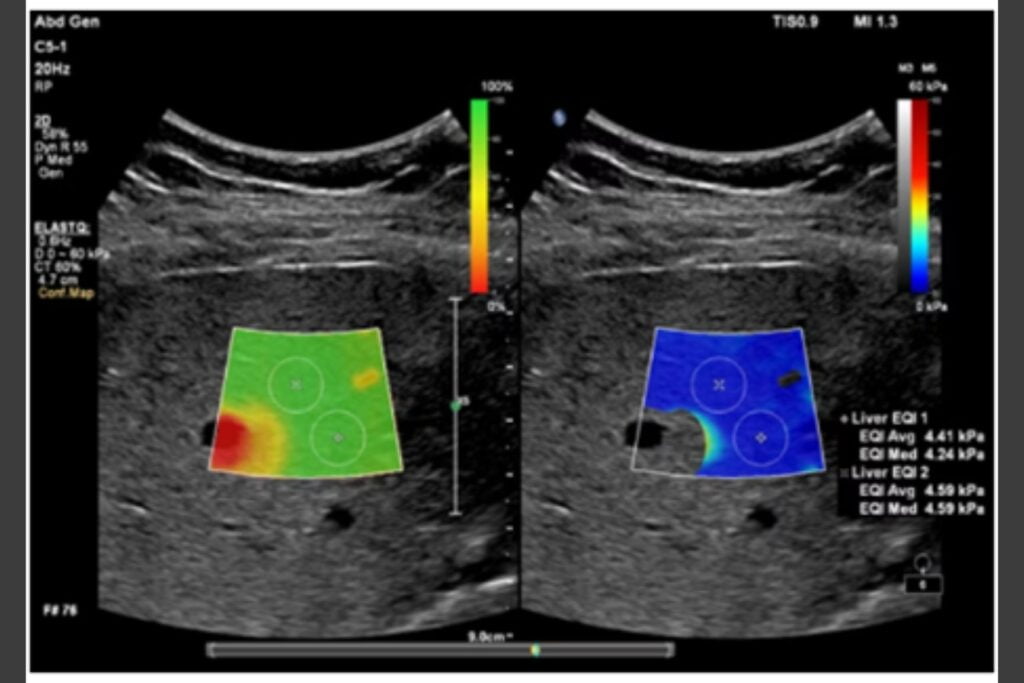
Elastography
Elastography is a medical imaging technique that is used to assess the elasticity or stiffness of tissues within the body.
It is a non-invasive method that provides additional information beyond what traditional ultrasound or other imaging modalities can offer. Elastography is particularly useful in the diagnosis and monitoring of various medical conditions.
- GENERAL QUESTIONS
Frequently asked questions.
Ans: An ultrasound is a medical imaging technique that uses high-frequency sound waves to create images of the inside of the body. It works by sending sound waves into the body and recording the echoes as they bounce back.
Ans: Ultrasounds are used to visualize various organs and tissues in the body, monitor pregnancies, diagnose medical conditions, and guide medical procedures. The doctor may recommend an ultrasound to assess specific concerns.
Ans: Preparation instructions can vary based on the type of ultrasound you need. In some cases, fasting or drinking water before the exam may be necessary
Ans: Ultrasound imaging is generally painless and non-invasive.
Ans: The duration of an ultrasound can vary depending on the area being examined
Ans: Ultrasound is considered safe, as it does not use ionizing radiation like X-rays. There are usually no known risks to patients undergoing ultrasound examinations.
Ans: Depending upon the scan, the radiologist may provide results immediately while in some cases immediate results are not given.
Ans: We allow patients to have a family member or friend present during the ultrasound for support.
Ans: Most ultrasounds may require a doctor’s prescription, while others can be scheduled directly with the center.
Ans: We offer various types of ultrasounds, including abdominal, pelvic, obstetric, vascular, and cardiac ultrasounds.
Ans: Ultrasound costs can vary based on the type and location of the exam. Insurance coverage also varies, so it’s essential to check with your insurance provider and the diagnostic center regarding costs and coverage.
Ans: We provide patients with a copy of their ultrasound images.